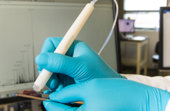
A team of engineers and scientists at the University of Texas have invented a pen that accurately identifies cancerous tissues during surgery.
The handheld instrument gives surgeons information on what tissue to cut and preserve in about 10 seconds.
“If you talk to cancer patients after surgery, one of the first things many will say is ‘I hope the surgeon got all the cancer out’, ” says Livia Schiavinato Eberlin, an assistant professor of chemistry at UT, Austin who led the team.
“It’s just heartbreaking when that’s not the case. But our technology could vastly improve the odds that surgeons really do remove every last trace of cancer during surgery.”
The current technique being used to determine the boundary between cancer and normal tissue is called frozen section analysis and it takes up to 30 minutes to prepare and interpret by a pathologist.
The MasSpec Pen was tested on tissues from 253 human patients and it was more than 96% accurate. The team expects to start testing the new technology during oncologic surgeries in 2018.
“Cancer cells have dysregulated metabolism as they’re growing out of control. Because the metabolites in cancer and normal cells are so different, we extract and analyze them with the MasSpec Pen to obtain a molecular fingerprint of the tissue,” says Eberlin.
When the MasSpec Pen completes its analysis, the words ‘Normal’ or ‘Cancer’ automatically appear on a computer screen. For certain cancers, such as lung cancer, the name of a subtype might also appear.
This research was accomplished by an interdisciplinary team, merging the fields of chemistry, engineering, and medicine.
The team and UT Austin have filed US patent applications for the technology and are now working to secure worldwide patents.
Copyright 2025 TheCable. All rights reserved. This material, and other digital content on this website, may not be reproduced, published, broadcast, rewritten or redistributed in whole or in part without prior express written permission from TheCable.
Follow us on twitter @Thecablestyle

